The sacroiliac joints are low motion synovial articulations formed where the ventral aspect of the iliac wing articulates with the fused transverse processes of the sacrum. The sacroiliac joints, in conjunction with substantial soft tissues, are responsible for transferring forces between the hindlimbs and axial skeleton. Allowing for the anatomical and biomechanical differences between bipeds and quadrupeds, many similarities between horses and humans exist with reference to sacroiliac joint pain. It is therefore prudent to take advantage of the relative abundance of human research on the subject. Similarities between the species include variable and often subtle clinical signs of sacroiliac joint region pain (Szadek et al, 2009; Barstow and Dyson, 2015; Maher et al, 2017), complex and inaccessible anatomy, presence of significant articular and periarticular degenerative or pathological changes seen in normally functioning individuals (Haussler et al, 1999; Eno et al, 2015) and the overlap in diagnostic imaging findings in painful and pain-free individuals (Archer et al, 2007; Thawrani et al, 2019). In humans and horses there is no reference or ‘gold-standard’ diagnostic test for sacroiliac joint pain (Szadek et al, 2009; Simopoulos et al, 2015) and in humans the issue is contentious. However, there is growing evidence indicating that a positive response to sacroiliac blocking is diagnostic in humans. In humans advanced cross-sectional imaging, such as computed tomography (CT) and magnetic resonance imaging (MRI), is undertaken to rule out other disease or injuries in the region (Thawrani et al, 2019). Equine veterinarians and human doctors similarly lack consensus regarding optimal treatment of individuals with sacroiliac joint pain, but substantial evidence supports sacroiliac joint injections with corticosteroids and physiotherapeutic interventions in human patients.
This topic is vast and cannot be covered in its entirety here. This article presents some of the key aspects of sacroiliac joint region pain in horses and addresses the challenges facing equine veterinarians when diagnosing and treating horses with suspected sacroiliac joint region pain.
There are numerous publications that should be reviewed should the reader seek greater detail on aspects of sacroiliac joint region pain including anatomy and morphology of the sacroiliac joints (Dalin and Jeffcott, 1986a, 1986b; Ekman et al, 1986), the clinical features of horses with sacroiliac joint region pain (Barstow and Dyson, 2015), the aetiopathogenesis and biomechanics of the sacroiliac region (Denoix, 1999, Goff et al, 2008) ultrasound of the pelvis and sacroiliac joint (Engeli et al, 2006; Head, 2014; Tallaj et al, 2019), sacroiliac injections (Denoix and Jacquet, 2008; Engeli and Haussler, 2012; Stack et al, 2016) and physiotherapy (McGowan et al, 2007; Clayton, 2012).
Anatomy, biomechanics and motion
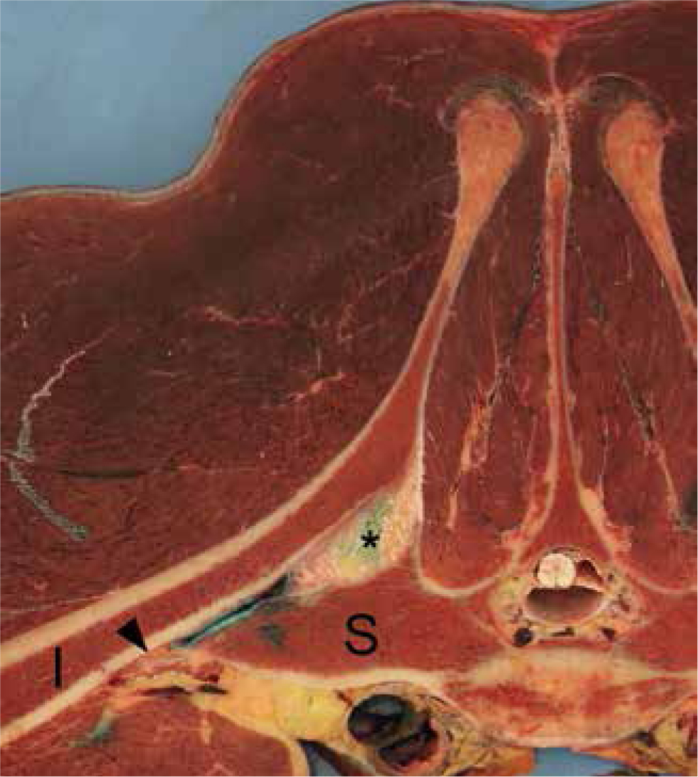
Sacroiliac joints are atypical diarthrodial joints, forming the articulation between the ventral aspect of the ilium and the sacral wing (Figure 1). The sacroiliac joint is angled at approximately 30° to the horizontal and is stabilised by the sacroiliac interosseous ligament, the dorsal (dorsal and lateral parts) sacroiliac ligament and ventral sacroiliac ligament. These ligaments blend with major fascial structures and ligaments of the back and pelvis including the thoracolumbar fascia and the sacrosciatic ligaments (Engeli et al, 2006). Numerous muscles have attachments to various parts of this fascial network, including the long head of the semimembranosus, middle gluteal and biceps femoris muscles (Goff et al, 2008). The sacroiliac joint is closely related to two sets of paired synovial articulations: the lumbosacral intertransverse joints cranially and lumbosacral dorsal articular process joints dorsomedially. The sacroiliac joint is the conduit for the propulsive forces generated by hindlimb action and is most likely subjected to shear forces during propulsion. The sacroiliac joints display a very limited and complex range of motion over approximately 2° compared to the adjacent lumbosacral junction that undergoes 10 times this range of motion (flexion/extension) (Degueurce et al, 2004, Goff et al, 2006). The latter is in part driven by contractions of the longissimus dorsi muscles inducing lumbosacral extension, and the contractions of the rectus abdominal muscles inducing lumbosacral flexion (Denoix, 1999). These muscles, as assessed by electromyography, are largely inactive at walk but fire twice per stride in trot (Denoix, 1999). This supports observations that there is significantly more skeletal excursion in all three planes in horses at walk compared to trot (Stubbs et al, 2006). The reduction in skeletal excursion, observed in trot, is thought to be a result of increased muscular stabilisation.
Epidemiology and pathology
The prevalence of sacroiliac joint region pain in the horse population is unknown, but degenerative changes in the sacroiliac joint are common in athletic horses (Dalin and Jeffcott, 1986a, 1986b; Haussler et al, 1999), with dressage, showjumping and eventing horses being over-represented in one clinical study of horses with sacroiliac joint region pain (Barstow and Dyson, 2015). In this same study 90% of horses with sacroiliac joint region pain had concurrent lameness, with many horses having concurrent diagnosis of hindlimb proximal suspensory desmopathy and/or forelimb foot related lameness (Barstow and Dyson, 2015). In humans, sacroiliac joint pain is considered part of low back pain. The number of people in the general adult population who experienced their first episode of low back pain in any one year was estimated at 6–15%, with as many as 30% of these patients reporting sacroiliac joint pain (Hoy et al, 2010; Rashbaum et al, 2016.
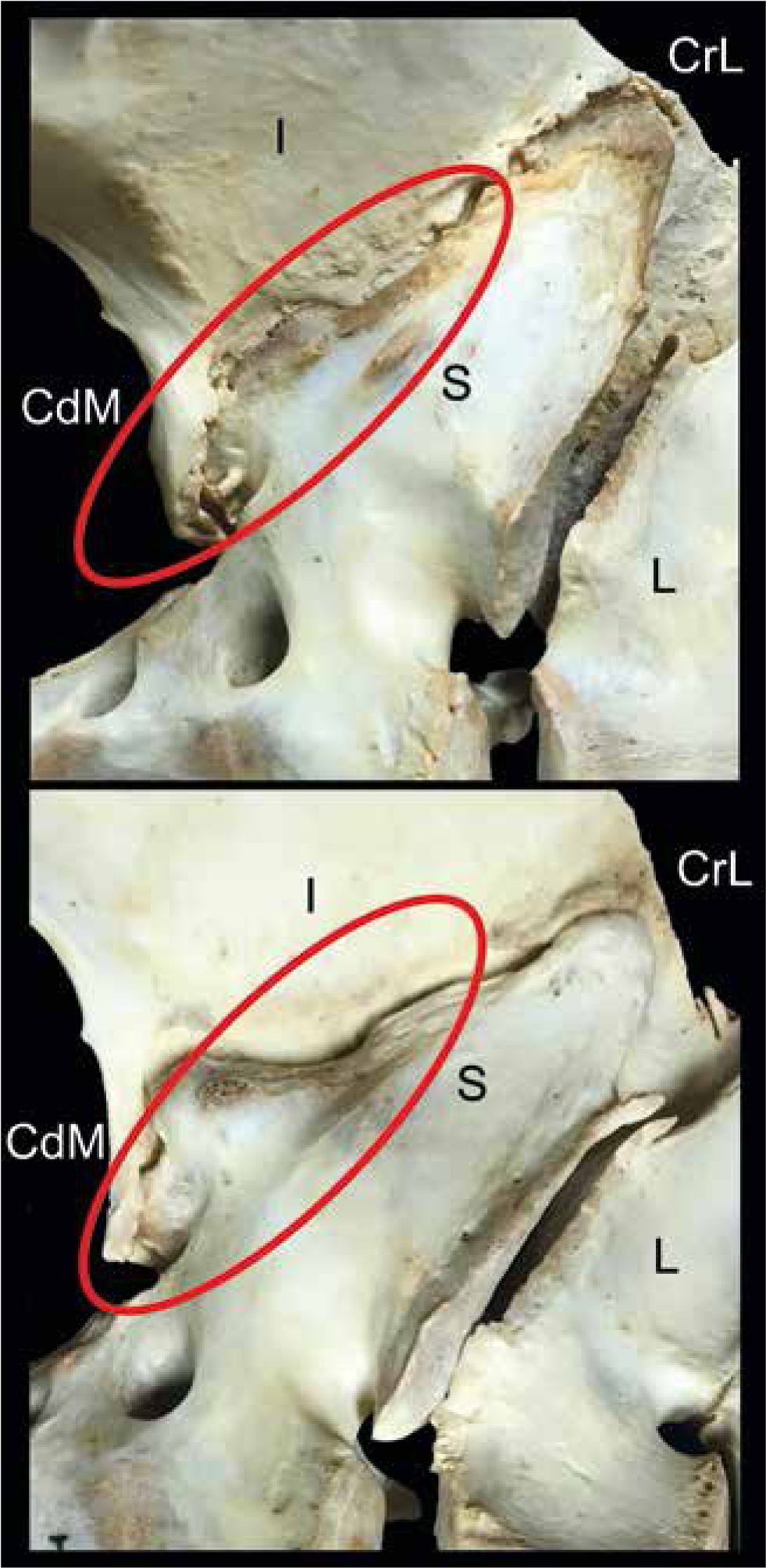
Degenerative changes in the sacroiliac joint have been documented in horses with and without clinical signs of poor performance or sacroiliac joint region pain (Jeffcott et al, 1985; Dalin and Jeffcott, 1986a, 1986b; Haussler et al, 1999). In one study, all 36 flat racing Thoroughbred racehorses (aged 4.5 years +/- 1.5 years) euthanised on the racecourse had sacroiliac degenerative lesions, and were graded as severe in one third of horses (Haussler et al, 1999). Most degeneration affected the caudomedial aspect of the sacroiliac joint. The sacroiliac articular surfaces of 20 of 41 asymptomatic horses were described as degenerate in another study (Dalin and Jeffcott, 1986a). The sacroiliac joints of 11 competitive horses with chronic poor performance attributable to sacroiliac joint region based on mild lameness, lack of impulsion, atrophy of the gluteal musculature and pain on palpation of the sacroiliac region had evidence of caudomedial periarticular bone modelling, but only two horses had sacroiliac joint arthrosis (Jeffcott et al, 1985). True arthrosis has also been noted in horses at post-mortem examination following diagnosis with chronic sacroiliac joint pain as determined by periarticular blocking (Nagy et al, 2020) and severe sacroiliac joint disease observed following chronic articular iliac fracture (Stubbs et al, 2010). It has been suggested that the periarticular bone modelling and caudomedial joint extension frequently observed in the sacroiliac joint are physiological adaptations in response to increased or cyclical loading, or from chronic instability resulting from dysfunction of the stabilising neuromuscular structures (Goff et al, 2008) (Figure 2). Asymptomatic humans had less movement through the SI joint compared to patients with sacroiliac joint pain supporting the thesis that SI joint disease and instability are connected (Nagamoto et al, 2015).
Diagnosis
Differentiating sacroiliac pain from thoracolumbar and lumbosacral pain is critically important so that appropriate treatment can be instituted. However, the latter is not always possible because of the close anatomical arrangement of these structures. Horses presenting for poor performance with signs of sacroiliac joint region pain must also undergo thorough screening of the appendicular musculoskeletal systems for concurrent lameness as this is very common (Barstow and Dyson, 2015).
Clinical presentation
Two studies have described the clinical presentation and diagnostic findings of horses with sacroiliac region pain (Dyson and Murray, 2003; Barstow and Dyson, 2015). The most common presenting signs were poor performance, not working well on the bit, with riders describing the horses as stiff in the back. In most cases for these horses the onset of signs was insidious. On the lunge horses lacked impulsion, became disunited in canter or cantered in a 4-beat fashion (Dyson and Murray 2003; Barstow and Dyson 2015). It is worth noting that in a study assessing back kinematics in lame horses, blocking of the lameness resulted in increased flexion, extension, lateral bending and rotation of the thoracolumbosacral spine increased in horses (Greve et al, 2017). This finding is interesting for several reasons. First, it highlights the importance of subjecting horses presenting for back stiffness to thorough examination of appendicular musculoskeletal systems. Second, it suggests that the biomechanics of the horse's back are altered in response to lameness, with the potential for development of back pain (Alvarez et al, 2007, 2008).
Visual assessment and palpation
Horses with sacroiliac joint region pain are often noted to have lumbar and hind quarter muscle atrophy and bony pelvic asymmetry (Jeffcott et al, 1985; Dyson and Murray, 2003, Barstow and Dyson, 2015). However, these signs are not specific for sacroiliac joint region pain as horses with facet pain and thoracolumbar impingement of the dorsal spinous processes can present in similar fashion (Girodroux et al, 2009). Pain on palpation of the gluteal and epaxial lumbar muscles and pain avoidance behaviours during manoeuvres designed to increase sacroiliac joint stress, known as sacroiliac provocation tests, have been noted in horses with sacroiliac pain (Haussler, 2003; Varcoe-Cocks et al, 2006). The accuracy of provocation testing to diagnose human patients with confirmed sacroiliac joint pain has been reviewed (Szadek et al, 2009) and pooled data from four studies showed good sensitivity (85%) and specificity (74%) when three or more sacroiliac provocation test results were positive. In horses a scoring system evaluating a number of clinical parameters including the presence of a plaiting gait when trotting, pelvic symmetry, muscle atrophy, response to palpation, and results of sacroiliac provocation testing has been described (Varcoe-Cocks et al, 2006). Cumulative scores were used to define horses as having mild, moderate or severe sacroiliac dysfunction (Varcoe-Cocks et al, 2006). However, the scoring system was not compared to response to diagnostic analgesia, cross-sectional diagnostic imaging or post-mortem examination.
Diagnostic imaging
When considering the interpretation of findings of diagnostic imaging of the sacroiliac joint and lumbosacral junction findings it is important to recognise the limited understanding of the clinical significance of the observed changes. Degenerative changes were commonly seen in the sacroiliac joint of human patients with no low back pain when undergoing diagnostic imaging (Eno et al, 2015; Thawrani et al, 2019).
Scintigraphy
Scintigraphy is a very commonly used imaging modality in the investigation of poorly performing and lame horses. The indications and limitations of scintigraphy have been reviewed (Archer et al, 2007). The review highlighted the challenge of interpreting the clinical significance of mild and moderate increases in uptake of radiopharmaceutical, and the risk of false positive diagnosis. A study compared scintigraphy to the final diagnosis in sport horses (n=480) with poor performance and/or lameness (Quiney et al, 2018). For all diagnoses, scintigraphy had a sensitivity and specificity for detection of injury of 48% and 94%, respectively. When used in the detection of sacroiliac joint region pain (based on blocking) the sensitivity and specificity were substantially lower and in another study by the same group there was no significant correlation between scintigraphy findings and periarticular sacroiliac joint blocking (Nagy et al, 2020). However, scintigraphy of this region is a useful screening tool for pelvic fractures and in horses not amenable to blocking (MacKinnon et al, 2015).
Ultrasound
Numerous descriptive studies illustrate the use of transcutaneous and transrectal pelvic ultrasound for the identification of muscle, ligament and bone injury in horses' lumbar, pelvic and sacral regions (Tomlinson et al, 2003; Kersten and Edinger, 2004; Engeli et al, 2006; Goodrich et al, 2006; Head, 2014; Boado et al, 2020; Nagy and Quiney, 2020). Ultrasound is indicated for the diagnosis of stress and complete fracture of the ilium (Pilsworth et al, 1994; Geburek et al, 2009), sacral fracture (Tomlinson et al, 2003), desmitis of the dorsal sacroiliac ligament (Tomlinson et al, 2003; Engeli et al, 2006), identification of periarticular enthesophyte formation along the ventral aspect of the sacroiliac joint (Barstow and Dyson, 2015; Tallaj et al, 2020) and lumbosacral disc disease or intertransverse joint periarticular modelling (Barstow and Dyson, 2015; Boado et al, 2020; Vautravers et al, 2020). Ultrasound has been used effectively to assess the cross-sectional area of the multifidus muscle, atrophy of which has been observed in horses with facet fracture or disease and sacroiliac joint pathological changes at post-mortem (Stubbs et al, 2010). There is no acoustic window for the dorsum of the sacroiliac joint.
Radiography
Techniques for the acquisition of high-quality radiographs have been described for the lumbosacral and sacroiliac region in horses (Gorgas et al, 2007, 2009). The technique was useful in identifying lumbosacral abnormalities such as transitional and hemi-transitional vertebrae, and for determining the outline of the sacroiliac joints. However, radiography for sacroiliac disease is not widely performed in horses, probably because of the necessity for general anaesthesia and the absence of diagnostic criterion for sacroiliac joint disease. Radiography is useful in standing horses for some pelvic fractures (acetabular, sacral, tuber coxae and tuber ischium) and should be considered if these injuries are suspected (Geburek et al, 2009).
Computed tomography
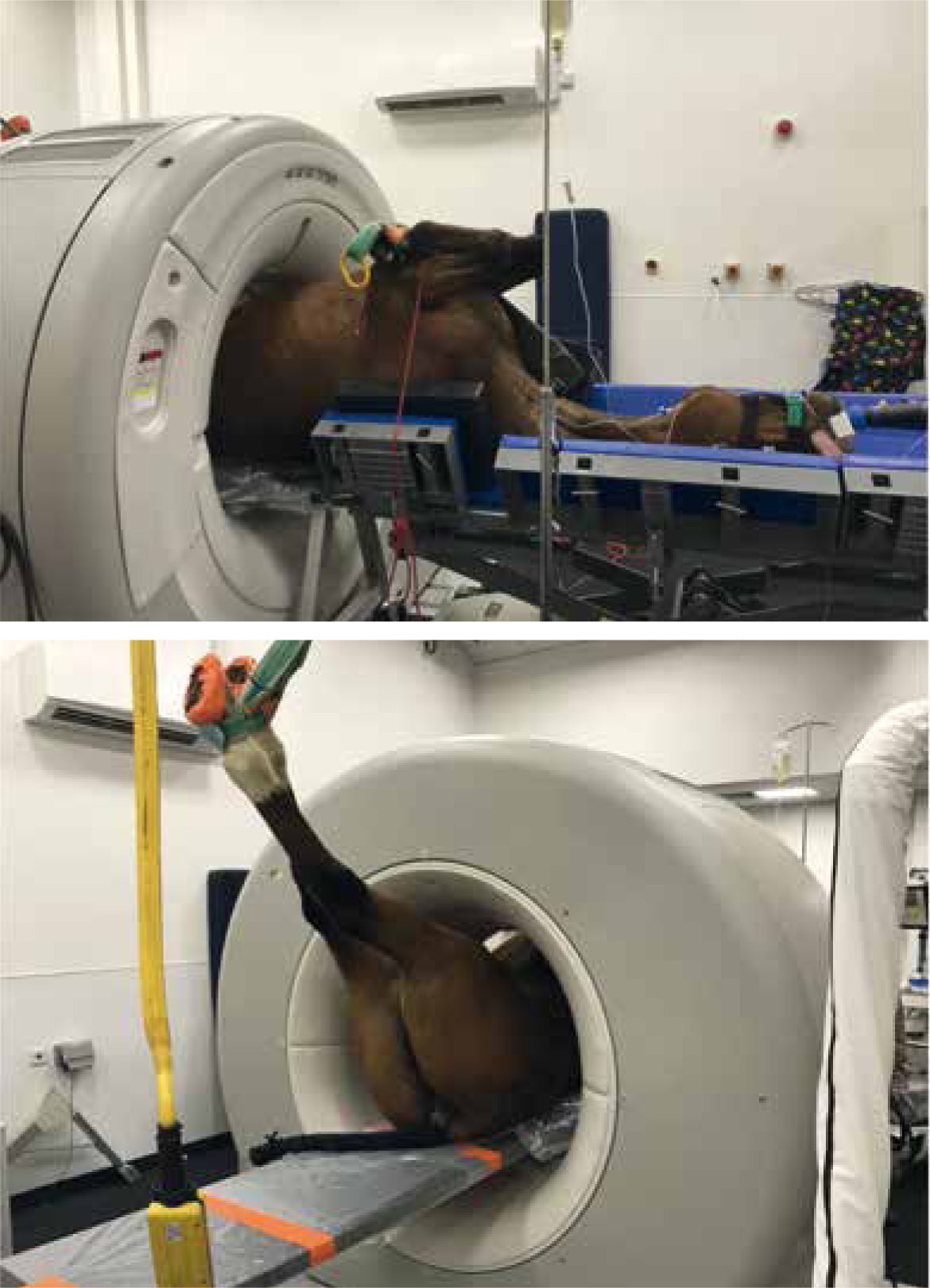
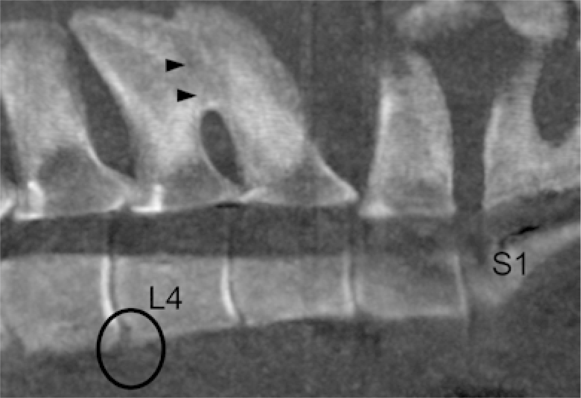
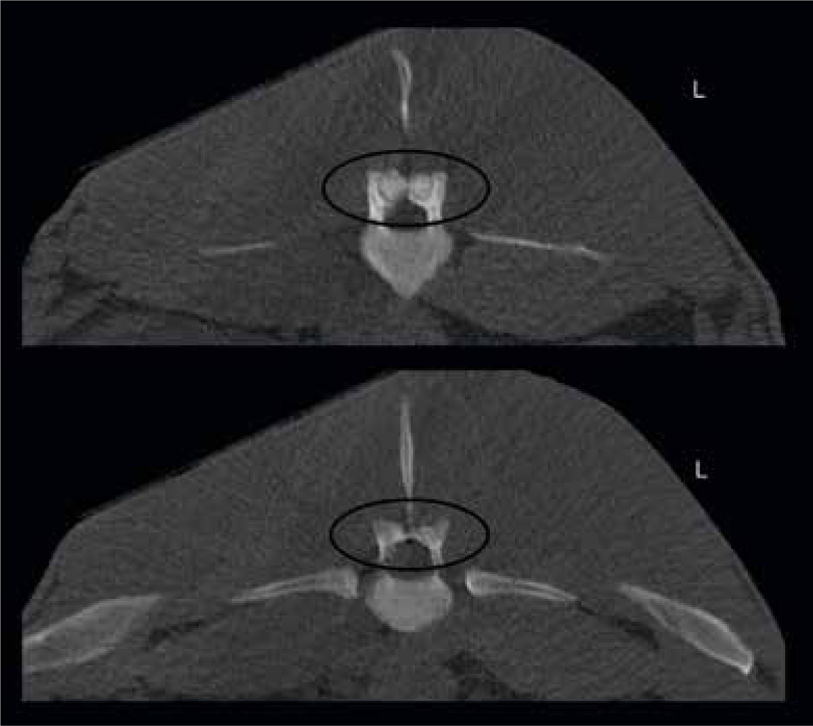
Computed tomography is very useful in the evaluation of human patients with low back pain to rule out fractures and congenital dysmorphism (Rashbaum et al, 2016). In humans, periarticular modelling and alterations in the thickness of the subchondral bone plate are routinely identified and measured; however, these changes are not predicative of response to intra-articular blocking (Eno et al, 2015; Thawrani et al, 2019). Post-mortem computed tomography performed on dissected lumbosacropelvic specimens identified lumbosacral disc disease and disc protrusion in one horse (Krueger et al, 2016) and a lumbar fracture in another horse (Tomlinson et al, 2003). With large bore (85–90 cm diameter) scanners, computed tomography imaging of the sacroiliac region in horses up to 525 kg is possible (Stack, 2019) and pelvic computed tomography is performed routinely in two equine clinics: the Philip Leverhulme Equine Hospital at the University of Liverpool, UK and the Lingehoeve Diergeneeskunde, in the Netherlands (Figure 3). Computed tomography represents a potentially useful tool for evaluating the osseous and articular structures of caudal lumbar, lumbosacral junction, pelvis and sacroiliac region with potential to identify impinging dorsal spinous processes (Figure 4), fused intertransverse joints, articular process (facet) joint disease or ankylosis (Figure 5), periarticular modelling and enthesopathy (Figure 6), fracture and dysmorphism. Acquisition and analysis of such imaging data will improve the understanding of disease prevalence and the significance of changes seen in this area. The biggest obstacles to scanning the sacroiliac region remains the lack of access to wide bore computed tomography scanners and the need to perform scans under general anaesthesia.

Sacroiliac injections
Owing to difficulties with interpreting diagnostic imaging changes, positive response in clinical signs following injection of local anaes-thesia targeting the sacroiliac joint is recommended as confirmatory of sacroiliac joint region pain in horses (Dyson and Murrary, 2003; Barstow and Dyson, 2015) and in humans (Szadek et al, 2009; Simopoulos et al, 2015). However, in contrast to humans sacroiliac injection techniques are not accurate for intra-articular injection of the sacroiliac joint (Engeli et al, 2004; Cousty et al, 2008; Stack et al, 2016). Injectate dispersion can be widespread particularly when large volumes (15–20 ml) of injectate are used (Engeli and Haussler, 2012; Barstow and Dyson, 2015), but this is also observed with smaller volumes (6 ml) (Stack et al, 2016). Pain in horses responding to sacroiliac targeted local anaesthetic injection therefore may be emanating from any of the sensitive structures of the sacroiliac joint or lumbosacral region. In humans the pain sensitive structures of the lower back have been identified as nerve roots, the intervertebral disc, dorsal and ventral vertebral ligaments, supraspinous ligaments, joint capsules of the dorsal articular process joints, interspinous ligaments, intra-articular synovial folds, the vasculature, periosteum, and muscles of the back (Maher et al, 2017). It is probable therefore that similar structures are sensitive in the horse, but the innervation has not been described.
An improvement of 50% in clinical signs is generally considered a positive test in horses but in human studies a positive response of 70-90% is considered positive (Szadek et al, 2009). In the most recent SI injection technique study (Stack et al, 2016) cranial parasagittal and caudomedial ultrasound-guided techniques were used to inject 6ml of iodinated contrast in cadavers and the location of the contrast was assessed by computed tomography scan. All injections were located within 2 cm of the sacroiliac joint margin but dispersal of contrast to other structures, including the joints of the lumbosacral junction, was common highlighting lack of specificity with injections targeting the sacroiliac joint in horses. Caudomedial injections were more difficult to perform and frequently resulted in contact between the injected solution and neurovascular structures, including the sciatic nerve. Consequentially, it is not recommended to inject local anaes-thesia via this approach.
There is not yet a validated diagnostic algorithm for sacroiliac joint region pain in horses. Rather diagnosis must be made based on clinical reasoning and interpretation of the clinical findings, including provocation tests, the results of diagnostic imaging, and the response sacroiliac joint region blocking. Other axial and appendicular musculoskeletal pathologies are frequently found in conjunction with sacroiliac joint region pain and should be ruled out.
Treatment
Selecting the appropriate treatment for horses with sacroiliac joint region pain is confounded by numerous factors including lack of specific diagnosis in some horses as discussed, and the paucity of literature reporting outcomes following treatment in horses. Additionally, many horses presenting with sacroiliac joint region pain have concurrent injuries and conditions that may require contradictory rehabilitation schedules. Before embarking on treatment every effort should be made to rule out concurrent orthopaedic injury, notably hindlimb proximal suspensory desmitis. There have been numerous reviews of treatment outcomes in humans indicating fair outcomes following non-surgical therapy (Simopoulos et al, 2015; Rashbaum et al, 2016; Al-Subahi et al, 2017; maher et al, 2017; Prather et al, 2020). Although clear consensus is lacking in humans the mainstays of non-surgical treatment are pain reduction with medication and sacroiliac joint injection by corticosteroids, cessation of inciting activity (Prather et al, 2020) and physiotherapy. Physiotherapy for sacroiliac joint pain in humans consists of combinations of manual therapy (manipulation), therapeutic exercise and application of external devices such as support belts (Al-Subahi et al, 2017; Prather et al, 2020). Other treatments such as therapeutic laser and kinesiology tape lack evidential support.
In horses with a specific diagnosis, for example, desmitis of the dorsal sacroiliac ligament, or fracture of the sacral spinous process, treatment should be directed towards that specific injury. In horses with sacroiliac joint region pain the fundamentals of treatment are closely aligned with those of human patients. This is achieved through systemic administration of non-steroidal anti-inflammatories, corticosteroid injection targeting the sacroiliac joint, and rest. Horses with muscle spasm may benefit from muscle relaxants such as methocarbamol, although it is not licensed for use in horses in the UK (Muir et al, 1992). Treatment aimed at reducing pain should be aggressive enough to allow early instigation of physiotherapy and a graduated controlled exercise programme, the ultimate aim of which is to strengthen core and hindlimb musculature. Paddock rest on flat firm ground will be suitable for most horses. Excessive stable rest can be deleterious to those horses where neuromuscular sacroiliac joint dysfunction is suspected, as rest may further exacerbate muscle weakness and sacroiliac joint instability.
The only report of corticosteroid injection targeting the sacroiliac joints in horses was published by Nagy et al (2020). In this study reporting outcome following diagnosis of sacroiliac joint region pain, 32 horses received corticosteroid injections targeting the sacroiliac joint but the outcome of horses receiving injection and those not receiving injections were not reported separately (Nagy et al, 2020) making it impossible to draw any conclusion about efficacy of sacroiliac injections in this cohort. Even in humans it is unclear if corticosteroid injection improves overall prognosis for patients with sacroiliac pain (Simopoulos et al, 2015), although positive short term results are noted frequently. One study in particular reported better outcomes following combined intra-articular and periarticular sacroiliac injection with corticosteroid compared to those receiving only intra-articular corticosteroids (31% vs 12.5% patients were pain free at 3 months post-injection) (Prather et al, 2020). This is note-worthy for veterinary practitioners as this is the likely distribution of corticosteroid following sacroiliac joint targeted injections in horses (Dyson and Murray, 2003; Engeli et al, 2004; Stack et al, 2016).
Rehabilitation
Effective rehabilitation in horses should consist of a combination of physiotherapy (manipulation and therapeutic exercises) (Clayton, 2012) and a controlled exercise programme (Cottriall et al, 2008). Early engagement with an Association of Chartered Physiotherapist in Animal Therapy (ACPAT) physiotherapist is strongly advised. All other therapists, including chiropractors and osteopaths, should be listed on the register of musculoskeletal practitioners (RAMP). Only eight horses (9.5%) of 84 horses diagnosed with sacroiliac joint region pain were treated by an ACPAT physiotherapist in a study reporting the outcome of horses with sacroiliac joint region pain (Nagy et al, 2020). Institution of exercise levels that might precipitate clinical signs of sacroiliac joint region pain should only commence when pain has been controlled. Core strengthening exercises have been endorsed by numerous authors in both human and equine fields (McGowan et al, 2007; Clayton, 2012; Prather et al, 2020). In horses these consist of baited stretches where the stretch is maintained by active contraction of thoracolumbar epaxial and abdominal wall musculature. The purported effect is improved stabilisation of the sacroiliac joint via stronger core musculature. Increased cross-sectional area (hypertrophy) of the multifidus muscle has been recorded in horses following baited stretching programmes (Stubbs et al, 2011). Therapeutic exercise targeted at important muscle groups (hamstrings, quadriceps and gluteals) are also considered essential to re-establish sacroiliac joint stability in weak horses (McGowan et al, 2007; Goff et al, 2008) and in humans this is achieved through closed kinetic chain strengthening exercises such as squats and lunges (Prather et al, 2020). In horses a similar effect might be achieved by walking over ground poles, but this has not been evaluated.
It is unclear which gait is most beneficial for rehabilitation during the controlled exercise programme in horses with sacroiliac joint region pain, but it stands to reason that canter should be avoided early on as the sacroiliac joint is asymmetrically loaded in canter (McGowan et al, 2007; Goff et al, 2008). In walk the horse's back is not fully stabilised by the longissimus dorsi or the transversus abdominus and undergoes greater excursion compared to trot (Denoix, 1999; Stubbs et al, 2006). This likely increases forces applied to the sacroiliac ligaments and may increase sacroiliac joint mobility. It is probable that mobility exercises such as manipulative therapies and walking exercise are advantageous in some horses with stiffness but increasing mobility in horses with weak or poor neuromuscular control should be avoided (Al-Subahi et al, 2017; Prather et al, 2020).
In human studies short term pain reduction was noted following manipulative therapy consisting of manually induced high frequency low amplitude forces applied through the sacroiliac joint region (Al-Subahi et al, 2017; Prather et al, 2020) but studies in horses are lacking and there are concerns about the repeatability of such techniques between individual therapists. At trot the horse's back is stabilised as a result of increased core muscular activity. This gait therefore represents the best gait for strengthening these muscles and should be used as part of the graduated controlled exercise programme. Lunging exercise in trot increased the electromyographic activity in the longissimus dorsi indicating that lunging should also form part of the exercise regimen once substantial muscle strength has been restored. Side reins did not appear to increase longissimus activity in the same study (Cottriall et al, 2008), indicating that such equipment may not be useful for this purpose. Treadmills, both land and water, have been recommended for rehabilitation of horses with back pain but close supervision by experienced practitioners is advised (Nankervis et al, 2017). Other treatments such as extracorporeal shockwave therapy and therapeutic lasers have also been used to treat back pain but lack evidential backing. Horses should receive regular examination by veterinarians and ACPAT physiotherapists or other suitably qualified therapists (RAMP) to ensure the horse is not developing signs of SI joint region pain and to amend the graduated controlled exercise programme and physiotherapeutic programme.
Overall, the prognosis for horses with sacroiliac joint region pain is considered fair. Frequently, horses return to lower-level activity following diagnosis and treatment of sacroiliac joint region pain. Only eleven (13%) of 84 horses diagnosed with sacroiliac joint region pain remained in full athletic function for 1 year following rehabilitation (Nagy et al, 2020,) but a further 27 (32%) horses returned to ridden work but at a lower level of activity.
Conclusions
Borrowing heavily from the human literature allows equine practitioners to make sensible assumptions about diagnosis and treatment of sacroiliac joint region pain, but many aspects of this challenging disease still remain poorly understood in both humans and horses. Sacroiliac joint region pain is a complex disease, for which there is no validated diagnostic algorithm in horses. Substantial research is warranted in order to establish the significance of lumbosacral junction as a potential source of pain, a region that has received little attention, and lies in close proximity to the sacroiliac joint. The advent of large bore computed tomography scanners is enhancing the ability to examine the osseous anatomy of the lumbosacral and sacroiliac regions in some horses and identify facet joint disease, impingement of transverse and dorsal spinous processes, periarticular modelling and spondylosis. Currently a high of degree of suspicion of sacroiliac joint region pain can be assumed in horses that are performing poorly, lack impulsion, have back or hind quarter muscle atrophy, are positive to sacroiliac provocation testing, and show at least modest improvement to sacroiliac joint region blocking, under the proviso that the lower limbs have been thoroughly investigated. Treatment options in horses are limited but the combined strategy of pain control and early institution of therapeutic exercises such as baited stretches and a graduated controlled exercise programme, once pain has been resolved, should be implemented. Active engagement with ACPAT physiotherapists should be encouraged.
KEY POINTS
- In horses and humans, diagnosis of sacroiliac joint pain is controversial, but in humans 75% reduction in pain following sacroiliac joint blocking is considered to be diagnostic for the condition.
- This is based largely on the fact that sacroiliac injections in humans (fluoroscopically guided) are highly accurate for injection of the SI joint itself, whereas in horses ultrasound-guided sacroiliac injections are periarticular, leading to a lack of specificity in horses.
- A ‘gold standard’ diagnostic test for sacroiliac joint disease does not exist in horses making prescription of treatment and interpretation of response to treatment challenging.
- In humans advanced imaging such as magnetic resonance imaging and computed tomography is important in ruling out other lower back injuries or conditions but are not highly accurate for identifying patients with sacroiliac joint pain. Similarly, it is very important to rule out other conditions affecting the caudal spine and pelvis in horses, but this can be difficult as cross-sectional imaging of lumbosacral and sacroiliac regions are not routinely performed.
- Treatment of horses and humans with sacroiliac joint region pain consists of reducing pain with rest, non-steroidal anti-inflammatories and sacroiliac targeted corticosteroid injections, and increasing neuromuscular stabilisation through therapeutic exercises and controlled exercise. The evidence for these treatments in horses is not derived from clinical trials but from extrapolation from experimental studies, human trials and clinical experience.
- Prognosis for horses with sacroiliac joint region pain appears to be fair.


