

Nursing care of horses with ocular disease is an integral part of their veterinary treatment. This should include a holistic approach to their care, from pain assessment to nutrition, using subpalpebral lavage catheters and helping manage potential behavioural issues.
Recognition of ocular pain
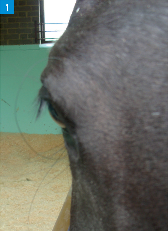
Ocular disease, particularly corneal diseases, can result in significant discomfort. While the pain level of the horse with his eye clamped shut who will not allow you anywhere near him is easy to assess, more subtle signs might be missed. These can be important, especially when assessing the response to treatment. As far as possible, the level of ocular pain should be assessed from outside the stall. In animals wearing protective eye masks, these should be removed and the horse left undisturbed in the stall for a short period of time to better assess comfort. The angle of the upper eyelashes is an excellent indicator of ocular pain (assuming that motor function to the lid is normal). Horses with ocular pain will tend to have the eyelashes angled down (Figure 1) (Gilger and Stoppini, 2005). Rather than relying on a descriptive assessment of ocular pain, which can vary between observers (for example, ‘very painful’ versus ‘mild discomfort’), a semiquantitative scale can be developed which includes how open the eye is (for example, 80%, compared to normal) and the angle of the eyelashes (for example, close to 90° would be normal, so 45° would suggest ocular pain). A specific ocular pain score has been investigated in horses, which includes blepharospasm, and this may be useful if further studies validate the initial findings (Ortolani et al, 2021).
Helping horses deal with limited vision
Even if the ocular condition does not have a significant impact on vision, in many instances protective eye masks are used – this renders the horse blind in that eye when wearing the mask (Figure 2).
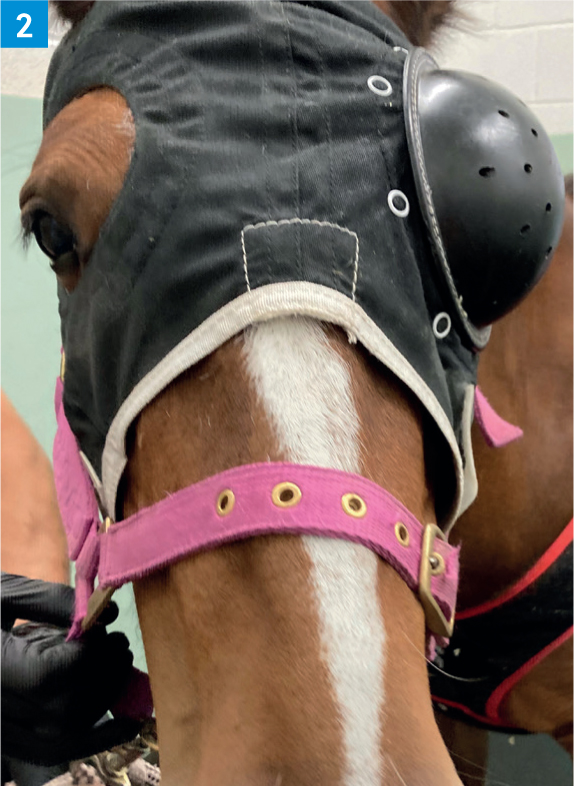
Most horses tolerate this well after a period of adaptation, but using voice cues is beneficial when approaching and working around these horses to ensure they are aware you are there. If vision is lost long term due to the eye condition, most horses are able to return to their previous ‘job’, including ridden work.
Subpalpebral lavage systems
A subpalpebral lavage system is an ophthalmic catheter that can be inserted through either the superior (upper) or inferior (lower) eyelid, allowing medications to be delivered into the tear film without having to manually open the lids to place the medications onto the surface of the eye (Dwyer, 2013). While most horses will tolerate short term eye medications, they quickly become masters of evasion. Subpalpebral lavage systems allow for frequent and long-term treatments to be administered with relative ease, although horses will still need to be managed to avoid behavioural problems associated with their use.
Subpalpebral lavage system: individual medications versus ‘preloaded’ catheters
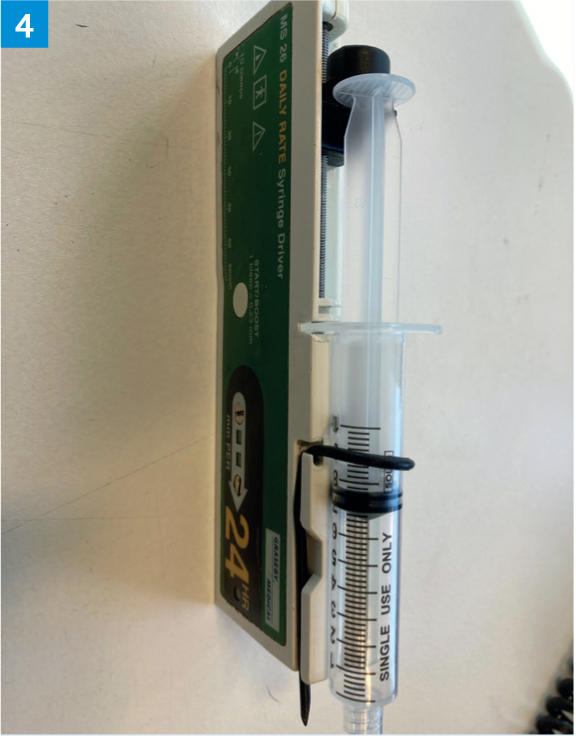
Administrations of medications individually, with flushing of the medication onto the ocular surface with air, rather than mixing of medications within the subpalpebral lavage system would be considered gold standard by many ophthalmologists for several reasons (Dwyer, 2013). Firstly, the effect of mixing on drug efficacy is largely unknown. Secondly, mixing medications essentially dilutes the concentration of each individual medication. However, horses tend to resent the air flushing more than the drugs themselves, and this method (both the air flushing and the individual application of medications) is very time-consuming, especially if multiple medications are being given at frequent intervals (a five minute delay is recommended between each medication to minimise the volume of liquid administered at each time (Dwyer, 2013)). In one study, the mixing of a variety of ophthalmic drug combinations did not affect antimicrobial efficacy in vitro for the majority of combinations (Johns et al, 2010). For these reasons, the author ‘preloads’ the whole tubing with individual aliquots of each medication and utilises a pump attached to the end of the subpalpebral lavage system, which allows for continuous administration of tiny volumes of medication onto the surface of the eye. The horses seem to tolerate this set up very well, it is much more time efficient and appears, at least anecdotally, to achieve similar outcomes. In the author's hospital, a pump made by Graseby Medical (Figure 3; 4) is used. The total volume of medications used in a 12 hour period is calculated (Drug A = Drug B + Drug C + Drug D, 0.2 ml each, given q2 hours = 4.8 ml). The scale on the front of the pump is then used to determine the rate in mm over a 24 hour period (as this is the time period pre-set into the pump). This number is then multiplied by 2 to give the rate for a 12 hour period. A 12 hour period is used as it limits the duration the drugs mix in the catheter and limits the volume of potentially expensive drugs which might be lost if the horse dislodges the pump unit.

Subpalpebral lavage system complications
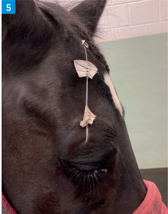
The subpalpebral lavage system can be placed in the upper eyelid or the lower eyelid based on clinician preference, the location of the lesion and the location of any eyelid issues which would interfere with placement. Several studies have investigated the incidence of complications comparing upper (Figure 5) and lower (Figure 6) lid placement, with the general consensus being that there is no significant difference between the two (Giuliano et al, 2000; Hays, 2013; Cornelissen et al, 2015; Quéré and Chahory, 2022). Whilst subpalpebral lavage systems undoubtedly make treatment of equine ocular disease far easier, there are some complications with their use, which should be monitored for. Most are relatively minor, but in some cases removal of the catheter may be necessary. The primary complications reported include iatrogenic corneal ulcer developing (Figure 7) if the footplate rubs on the cornea as a result of insufficient tension on or incorrect placement of the subpalpebral lavage system (Giuliano et al, 2000; Hays, 2013; Cornelissen et al, 2015; Quéré and Chahory, 2022). A sudden increase in discomfort may indicate this complication. Subpalpebral lavage system placement and tension should be checked on a daily basis to minimise the risk of this occurring. Palpebral (lid) cellulitis and abscessation can also occur (Giuliano et al, 2000; Hays, 2013; Cornelissen et al, 2015; Quéré and Chahory, 2022). In the author's experice, a small amount of lid swelling is expected within the first 24–48 hours after subpalpebral lavage system placement, but this should resolve quickly. Any other increase in swelling, especially if accompanied by heat or pain, could be caused by localised infection or in rare cases by regression of the catheter footplate so that medications are being delivered into the conjunctiva instead of onto the cornea. In most instances of lid swelling, local or systemic treatment is sufficient to resolve the condition, but in some cases, the subpalpebral lavage system will need to be removed. Growth of the conjunctiva around the footplate can potentially impede drug delivery or make removal of the catheter difficult (Quéré and Chahory, 2022). This is rare and tends to happen only when the subpalpebral lavage system has been in place for several weeks or months. In most cases, the subpalpebral lavage system can still be removed under standing sedation but in one report (Quéré and Chahory, 2022), removal under general anaesthetic was required. Another known complication is that medications may not be delivered onto the eye – if the catheter appears blocked (meaning medications cannot be pushed through, even with gentle pressure), it is likely to be blocked. Closely inspecting the line to check for any obvious areas of kinking or occlusion, or where medications might have precipitated can usually locate the source of the problem. Medications that are in suspension (such as voriconazole) appear anecdotally to be more likely to precipitate and occlude the subpalpebral lavage system although serum seems to be the culprit in some cases. In the author's experience, using serum from a different horse often solves this. If a blockage of a preloaded subpalpebral lavage system continues to occur, individual administration of medications followed by air to flush should be performed.


Behaviour
Many horses with ocular disease become long term residents because of the need for frequent treatment and re-examinations. Long-term hospitalised horses can become ‘sour’, especially if the only interactions they have with people involve treating their (often painful) eye. As such, it is important that as much time as possible is spent with these horses without treating their eye. This could involve regular grooming, owner visits, walking out in hand or simply going into the stable to greet the horse and then leaving. It is far better to establish this as ‘the norm’ than try to convince a horse in pain that not all humans are going to make their eye hurt. Counter conditioning, whereby a procedure or treatment is paired with giving some feed or treat in a structured way can also be beneficial as the horse starts to make a positive association with the procedure (Pearson, 2019). This will start to change the horse's emotional response to the procedure; they will perceive someone approaching their eye with drops in a more positive way. If they are feeling more positive, it has been shown that animals then have a greater tolerance of pain and discomfort (Wagner, 2010; Absi and Flaten 2016).
Colic
Horses hospitalised for eye conditions are at risk of developing colic for a number of reasons. In one study of 337 horses over a 12 year period, 72 (21.4%) developed colic signs, most of which were mild but a small proportion of which needed surgery (Patipa et al, 2012). The increased risk of colic is thought to be multifactorial, with contributing factors including the need for stall rest and resultant limited exercise and the administration of atropine, an anticholinergic drug, which may contribute to ileus (Hillyer et al, 2002; Ström et al, 2021). Chronic pain is also considered a risk factor as ocular conditions are commonly very painful due to the large number of nerve endings in the cornea. Pain is a known contributor to ileus in people and is likely to also contribute in horses (Patipa et al, 2012). Although not directly a cause of colic, non-steroidal anti-inflammatory drugs have the potential to mask gastrointestinal pain and thus a mild colic episode may go unnoticed, as is thought to occur in hospitalised horses which develop caecal impactions. Horses with painful conditions are also commonly on non-steroidal anti-inflammatory drugs for a prolonged period of time, increasing the risk of conditions such as right dorsal colitis, which can cause colic, oedema and loose faeces (Patipa et al, 2012).
Close observation of hospitalised eye cases is the best way to identify cases of colic before they become more serious. Subtle changes such as a decrease in appetite, increased time spent recumbent and decreased faecal output should be warning signs for a potential colic episode. If colic signs become more severe, a change in clinical parameters such as heart rate as well as an increase in a more generalized pain score may occur. As decreased exercise is considered a risk factor for colic, frequent hand-walking or turnout in a small, secure pen (if appropriate) may be beneficial (Hillyer et al, 2002).
Nutrition and body condition
Close attention should be paid to both the weight and body condition score (an assessment of overall fat covering) of ophthalmic cases. Weekly assessments of weight, either via a weigh tape or scales may be beneficial. Most hospitalised horses tend to gain weight if their appetite is good due to decreased activity, unless feed intake is limited accordingly (with owner permission). However, some horses will lose weight due to their poor appetite and these horses are often more challenging to manage. Hand grazing is a good way to improve appetite, and if necessary, grass can be picked for the horse to be fed in the stable. Trying different feeds may help, knowing that a fussy eater may like Food A one day and not the next. Gastric ulceration can contribute to a poor appetite in some horses, so gastroscopy and treatment as indicated may be beneficial (Sykes et al, 2015).
KEY POINTS
- Horses hospitalised for the treatment of ocular conditions require a holistic approach to their nursing care.
- Attention should be paid not only to treatment of the eye disease itself, but also to the horse's demeanour, general health and body condition.
- Using a subpalpebral lavage catheter can make treatment of chronic eye conditions much easier - learning how to troubleshoot these catheters is an important part of their management.
- Depending on clinician preference, medications can be given individually by the catheter, or via a continuous ‘pump’ system.